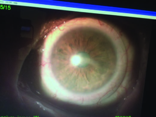
INDICATION: Corneal Scarring / Irregularity
This case illustrates that scleral shape rather than size and weight of the lens can sometimes be the major contributing factor to scleral lens decentration.
Do you know why some of your patients have poorly fitting scleral lenses?
Would you fit corneal lenses without measuring the cornea?
This 79-year-old male patient with central corneal scarring and irregularity OS due to recurrent herpes zoster. Multiple attempts to fit scleral lenses failed due to inferior scleral lens decentration.
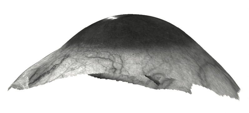
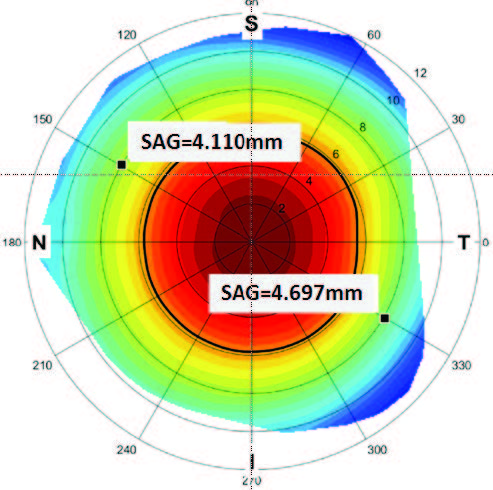
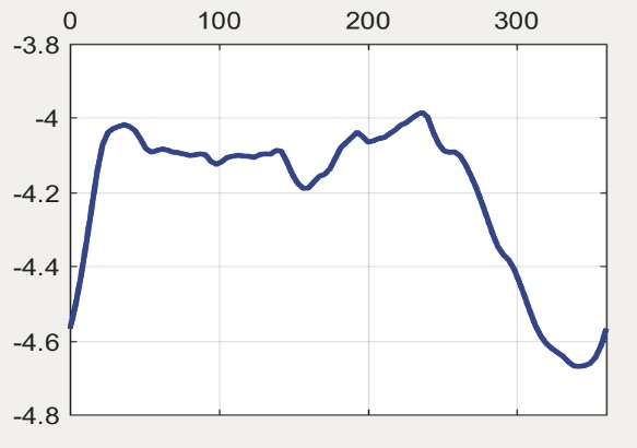
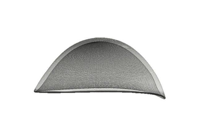
sMap3D™ ocular surface imaging (Figure 1) revealed a substantially steeper area inferotemporal at 330° than 180° away superonasally at 150°. The difference in the 2 areas can be quantitated using the SAG map (Figure 2). In this case, the SAG at an 8mm radius (16mm diameter) from the corneal center was 4.697mm at 330° and 4.110mm at 150°. The change in the scleral elevation pattern was best observed on the scleral shape plot which graphs the SAG value on the Y-axis vs. meridian on the X-axis at any diameter from the corneal center; in this case 16mm (Figure 3). The SAG values were relatively uniform throughout with the exception of a large depression (steep area) of over 500μ in depth centered at about 330° and about 100° wide. This scleral toricity plot was used to obtain quantitative information to manufacture a multi-meridian design conforming to this specific eye (Figure 4).
The resulting lens (Figure 5) was the first scleral lens designed for this patient that was comfortable and well centered; BCVA was 20/30.
Courtesy of Boaz Schwartz, OD, FAAO
